Dental bone loss is a progressive condition that can compromise tooth stability if left untreated. Early detection and proper management of bone loss play a critical role in maintaining dental safety and stability. So, can a dental bone loss X-ray detect this condition effectively? Let’s explore the detailed answer in the article below.
How X-Rays detect dental bone loss?
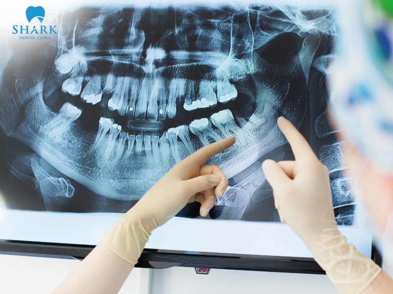
Dental bone loss often progresses silently beneath the gum line, making it difficult to detect with the naked eye. A dental X-ray allows dentists to assess the density and height of the alveolar bone surrounding the tooth roots. Based on the X-ray images, clinicians can identify the location of bone resorption, the extent of bone loss, and its severity.
When the jawbone is healthy and has a high mineral content, it effectively blocks X-rays, resulting in clear and well-defined images on the radiograph. In contrast, in cases of jawbone loss, X-rays pass through more easily, creating visible gaps or voids on the film.
By analyzing these differences, dentists can accurately diagnose and evaluate the degree of dental bone loss. This information enables them to develop an appropriate treatment plan to manage and improve the condition, ultimately helping to restore both chewing function and dental aesthetics for patients.

>>> See more: Can you get dental X-rays while pregnant?
What does bone loss look like on a dental X-ray?
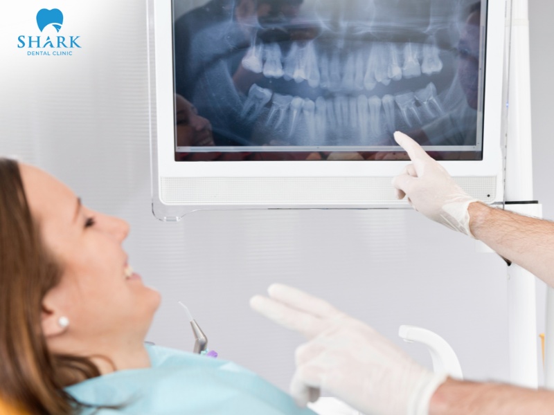
Many patients worry about how bone loss appears on dental X-rays. On a radiograph, a healthy jawbone typically appears as a bright white (radiopaque) area surrounding the tooth roots, with the bone margin located approximately 1–2 mm from the neck of the tooth.
In patients experiencing jawbone loss, the X-ray appearance varies depending on the severity of the condition, as outlined below:
- Horizontal bone loss: On the X-ray, the bone margin gradually lowers along the length of the tooth roots, resulting in a wider distance between the tooth neck and the bone crest compared to normal.
- Vertical bone loss: Bone resorption forms deep, angular defects that are clearly visible on dental X-rays. This is a typical sign indicating a localized and potentially serious infection, often prompting concerned patients to ask, “Is this an abscessed tooth infected root canal X-ray?”
- Furcation bone loss: In multi-rooted teeth affected by bone loss, radiolucent shadows can be seen on the X-ray. This indicates that the damage has extended into the space between the roots and requires early intervention.
Additionally, the loss of the lamina dura surrounding the tooth root may appear as radiolucent areas on X-ray images. This finding serves as a warning sign of inflammation and the breakdown of structural connections between segments of the jawbone. Because this type of bone destruction can look quite alarming on a monitor, it often prompts concerned patients to ask, “What does cancer look like on dental X-ray?” While oral malignancies can also present as destructive radiolucent lesions, your dentist is expertly trained to differentiate them from these common, localized infections.
>>> See more: How often should you get dental X-Rays?
Treatment options after bone loss diagnosis
For patients managing their care budget, researching the dental X-ray cost in Vietnam is often the first step in this diagnostic journey. Once the severity and specific condition of bone loss have been identified, the dentist will create a treatment plan based on the patient’s overall oral health. The primary goals of treatment are to halt further destruction of the jawbone, promote bone regeneration, and restore normal dental function.
Here are some common treatment options that may be recommended:
- Scaling and root planing: To prevent further progression of bone loss, dentists perform thorough cleaning beneath the gum line, removing tartar, plaque, and harmful bacteria.
- Bone grafting: In certain cases, autogenous bone or synthetic bone graft materials may be used to fill areas affected by bone loss. These grafts provide a stable foundation for natural teeth or prepare the site for dental implant placement.
- Bone regeneration procedures: Dental barrier membranes are applied to prevent external factors from invading the bone-defect area, creating a favorable environment for natural bone growth and stimulating tissue regeneration.
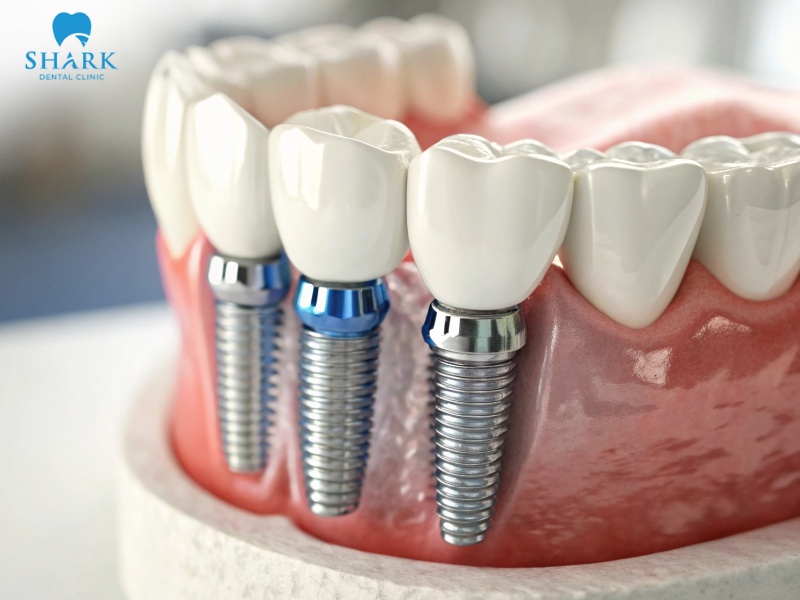
- Dental implant placement: In cases of severe, irreversible bone loss leading to tooth loss, dental implants may be recommended. This solution restores chewing function, improves aesthetics, and helps prevent further jawbone resorption.
Using a dental bone loss X-ray is a necessary diagnostic step that supports accurate assessment and effective treatment planning. If you notice signs such as loose teeth, swollen or reddened gums, or bleeding gums, seek dental care and undergo an X-ray examination promptly to receive timely and appropriate treatment!
>>> See more: Dental cavity X-ray