
Dental cavities often begin as tiny spots in the spaces between teeth or near the tooth roots—areas that are difficult to observe with the naked eye. So, can a dental cavity X-ray detect tooth decay effectively? The article below will help answer this question while also providing detailed information about dental X-ray techniques used in dentistry.
What a dental cavity x-ray reveals about tooth health?
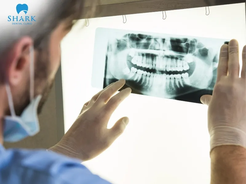
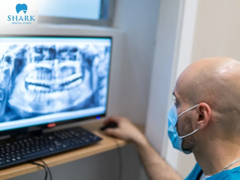
A dental X-ray helps dentists detect tooth decay through detailed imaging. By analyzing the enamel layer, the extent of demineralization, and how X-rays interact with the mineral density of tooth structures, accurately evaluate the presence and extent of tooth decay.
When tooth decay begins, mineral density decreases, causing the tooth structure to become thinner and weaker. As a result, cavities appear on X-ray images as dark areas or shadowed zones, which are clearly distinguishable from healthy tooth tissue. These visual signs are crucial for identifying the presence, location, and severity of cavities, allowing dentists to develop effective and safe treatment plans.
Physical symptoms that signal the need for an X-Ray
Several clinical symptoms of tooth decay indicate the need for a dental X-ray to confirm the diagnosis, including:
- Changes in enamel color: The appearance of chalky white spots or small brown discolorations on tooth surfaces or between teeth is an early sign of decay.
- Persistent tooth sensitivity: Ongoing discomfort, particularly when consuming hot, cold, or sugary foods and drinks.
- Toothache during chewing: Pain or discomfort when biting or applying pressure to the teeth.
- Swollen or reddened gums: This may signal infection or advanced decay within the oral cavity.
In these cases, X-ray imaging is essential for early detection and accurate diagnosis, helping dentists plan timely treatment and reduce the risk of infection or complications. If you are wondering, “Are dental X-rays safe?”, rest assured that modern digital radiography utilizes extremely low doses of radiation, making this crucial diagnostic step both secure and highly effective.
Treatment options after a confirmed cavity diagnosis
Once tooth decay is confirmed through a dental cavity X-ray, dentists will recommend appropriate treatment options based on the severity of the condition:
- Enamel remineralization: This is suitable for early-stage cavities. High-concentration fluoride is applied to replenish lost minerals, helping the enamel repair itself and halt the progression of decay.
- Dental fillings: Small cavities affecting the enamel or dentin can be treated with tooth-colored filling materials that restore function and aesthetics while preventing further decay.
- Dental crowns: Recommended for larger cavities where the pulp is not yet infected, crowns provide strength, durability, and aesthetic restoration.
- Root canal treatment: This is necessary when decay has spread to the pulp or tooth apex. After cleaning and sealing the root canal, a crown is placed to restore the tooth’s appearance and function.
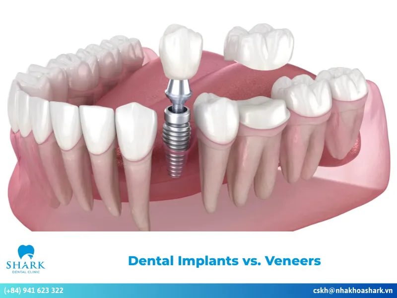
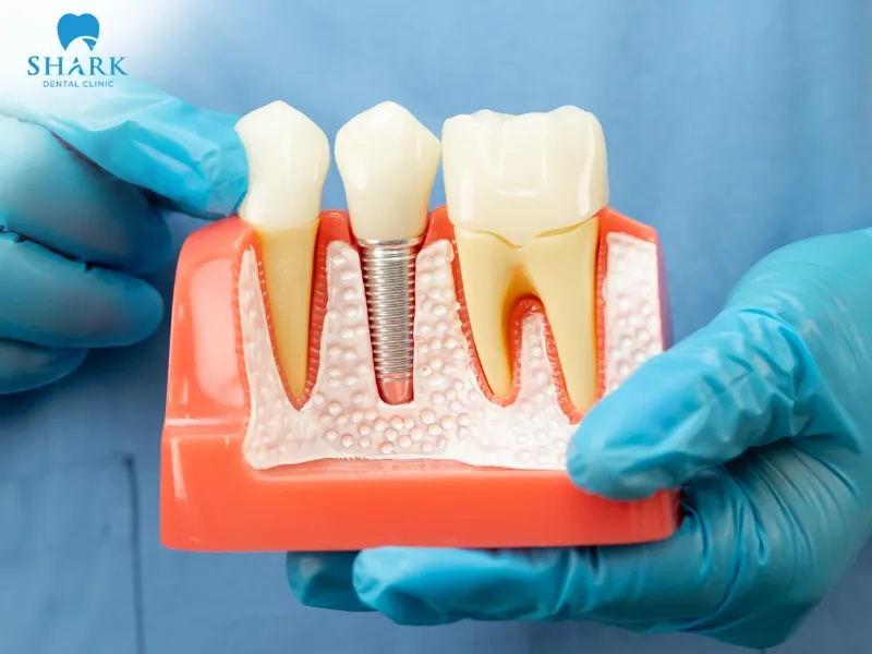
- Tooth replacement: If decay has completely destroyed the tooth and extraction is unavoidable, dental implants may be considered. Implant posts offer long-term durability and effectively replace missing tooth roots.
The choice of treatment depends on the specific oral health conditions and the severity of the cavity. Based on X-ray findings, dentists will guide patients toward the most suitable solution.
How tooth decay appears on X-Rays?
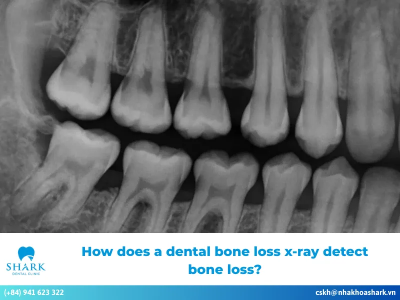
In dental X-ray images, tooth decay is identified by variations in brightness and density within hard tissues. Enamel and dentin are highly mineralized and block X-rays effectively, appearing bright white on the image.
In contrast, a fundamental part of learning how to read a dental X-ray is recognizing that areas affected by decay show up as dark or shadowed regions, indicating mineral loss caused by bacterial activity. Even subtle color changes on a dental X-ray allow dentists to detect early decay, evaluate its progression, and determine the most effective treatment approach to preserve tooth health.
Radiolucent vs. Radiopaque findings in cavity detection
In dental X-ray imaging, radiopaque areas represent solid, dense structures with a high mineral content, appearing bright white on the film. In contrast, radiolucent areas indicate hollow or low-density structures, with tooth decay showing up as shadowed zones or dark spots.
The contrast between radiopaque and radiolucent regions is crucial for dentists to assess and determine the severity of cavities. If a radiolucent area extends to the pulp chamber, it indicates advanced decay affecting the dental pulp, necessitating early intervention to prevent serious complications. Beyond decay, patients reviewing their scans often anxiously ask, “What does cancer look like on dental X-ray?” While less common, these serious pathologies can also present as irregular radiolucent or mixed lesions that disrupt normal bone architecture, further highlighting the importance of a comprehensive radiographic evaluation.

Can a dental cavity x-ray miss early or hidden decay?
While dental X-rays are considered effective and accurate diagnostic tools, they can sometimes miss cavities in their very early stages. Minimal mineral loss may not create sufficient contrast on X-ray images for clear detection.
Additionally, in patients with dental crowns or fillings, these restorative materials can block X-ray radiation, making it more challenging to identify early or hidden decay. Therefore, dentists often combine X-ray imaging with clinical examinations and their professional experience to diagnose and treat tooth decay accurately.
Preventive strategies to stop cavities before they develop
Tooth decay is a common dental condition that affects not only aesthetics but can also lead to serious oral health complications. The following preventive measures can help reduce the risk of cavities safely and effectively:
- Maintain proper oral hygiene: Brush your teeth at least 2-3 times daily with a soft-bristled toothbrush. Use toothpaste containing fluoride, which can help remineralize enamel and prevent cavities.
- Use dental floss and water flossers: Combining traditional flossing with water flossers enhances plaque removal between teeth, limiting bacterial buildup.
- Rinse regularly: Rinse your mouth with diluted salt water or a specialized antibacterial mouthwash.
- Adopt a balanced diet: Ensure adequate nutrition while limiting fast food, fried foods, and oily meals. Reduce consumption of foods high in sugar, acid, and starch.
- Schedule regular dental check-ups: Visiting a dental clinic every 3-6 months helps prevent and detect early signs of cavities. Whether you are looking for a routine exam or a precise dental x-ray in Ho Chi Minh, choose a reputable dental clinic equipped with modern technology and experienced dentists.

In summary, utilizing a dental cavity X-ray is a crucial diagnostic step that enables dentists to create accurate and effective treatment plans. We hope this article has provided you with a clearer understanding of dental X-rays and practical ways to care for and protect your oral health effectively.