
The advancements of modern dentistry have made it possible to diagnose and support the treatment of many oral conditions. And one of the common questions many people have today is whether an X-ray can detect cancer, and what does cancer look like on dental X-ray. Let’s explore the information in the article below!
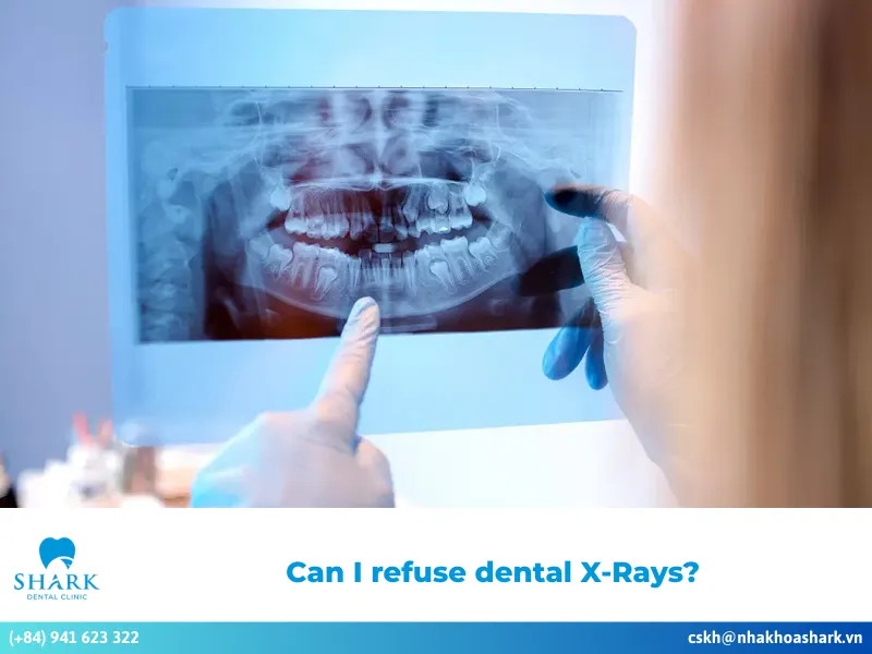
Can dental X-rays show cancer?
Dental X-rays can be helpful in the early detection of cancer in certain cases. While their primary purpose is to identify early tooth decay and assess bone health, dentists acknowledge that X-rays also play a crucial role in spotting cancer early on.
- Screening for abnormal issues: Dental X-rays primarily focus on bones and teeth, but they can also identify abnormalities in surrounding soft tissues. This capability increases the likelihood of detecting cancer at an early stage, leading to more effective treatment outcomes.
- Detecting early signs: Although dental X-rays are not standalone diagnostic tests for cancer, dentists can spot unusual changes in the cells of the oral cavity through X-ray images. This allows them to make an initial assessment of your health condition.
- Clinical examination: Dental X-rays are often used in conjunction with more specialized tests to evaluate your overall oral health, which is vital for detecting oral cancer.
- Monitoring changes in dental structure: X-rays enable dentists to observe concerning changes in the structures of the teeth and jaw. These changes can serve as warning signs for potential cancer.
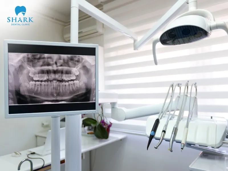
- X-Ray screening: Particular panoramic X-rays are significant in oral cancer screening. Opting for a dental x-ray in Ho Chi Minh provides a broad and relatively accurate view that helps dentists assess suspicious areas within the oral cavity. When planning your visit, the Dental X-ray cost in Vietnam is highly competitive, typically ranging from $5 to $15 (approx. 150,000 – 350,000 VND) for a panoramic film, while advanced 3D CT Cone Beam scans may cost between $20 and $40.
>>> See more: What OSHA standard applies to all medical and dental offices that have X-ray machines?
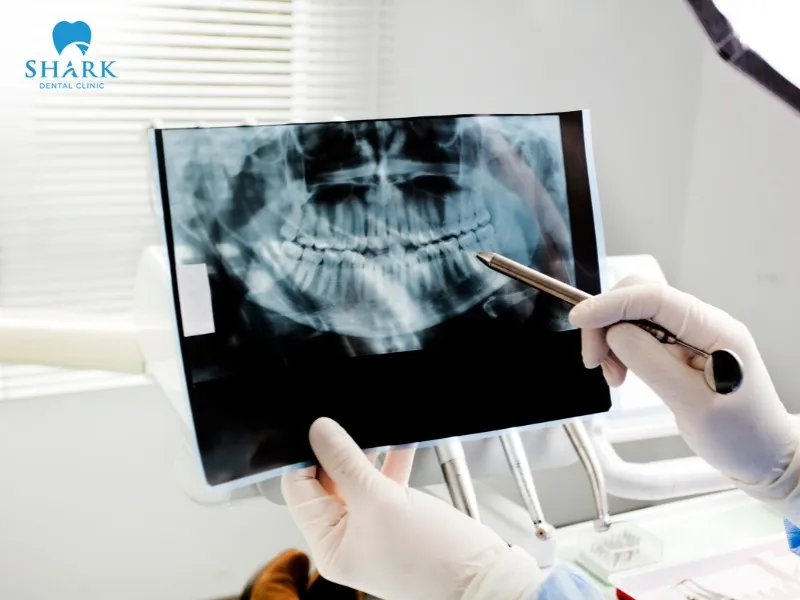
What does cancer look like on dental X-ray?
Dental X-rays allow dentists to observe gaps, hidden tooth structures, and more. They can help detect certain jaw and nearby bone cancers at an early stage. When learning how to read a dental X-ray for potential pathologies, specialists look for specific indicators of malignancy, as cancer often presents unique visual cues:
- Oral cancer: X-ray images may reveal changes in the bone. If oral cancer spreads to the jawbone, it may appear as dark areas indicating bone destruction or loss. Tumors can also distort the outline of the jawbone.
- Salivary gland tumors: These tumors can be detected on dental X-ray films, often appearing as dark regions accompanied by changes in bone structure and bone loss. In some cases, teeth may be displaced from their original positions, which can also be visible on X-rays.
- Jawbone tumors: X-ray imaging can help detect jawbone tumors early. These tumors may show up as both light and dark areas, causing the bone to expand or change shape in the jaw. The appearance of the tumor can vary based on its severity.
Limitations of dental X-Rays in detecting oral cancers
Despite their significant value in early cancer detection, dental X-rays have limitations when diagnosing oral cancer, particularly in its initial stages.
- Limited field of view: Dental X-rays primarily visualize hard tissues such as teeth and bone. However, oral cancer often begins in the soft tissues, meaning that X-rays cannot detect cancer in its early stages or identify subtle changes within those soft tissues.
- Image quality: The detail provided by X-ray films is often insufficient for diagnosing or accurately assessing cancer. Factors like exposure settings and image resolution can significantly affect a dentist’s diagnostic capabilities.
- Sensitivity of the film: The sensitivity and specificity of dental X-rays can vary in different situations. This indicates that X-rays serve only as an indirect method for diagnosing cancer.
Dental X-rays are not standalone diagnostic tools for cancer; they must be used alongside clinical evaluations and more advanced testing. Therefore, it is essential to maintain regular check-ups and comprehensive oral health evaluations to detect abnormalities early and address them effectively.
>>> See more: Dental care services in Vietnam
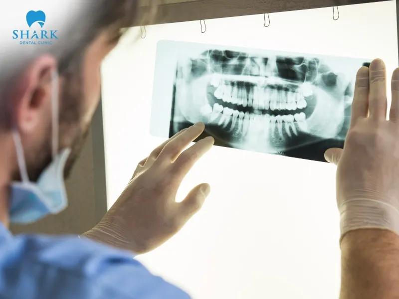
What do dentists do if they suspect cancer?
If a dentist identifies potential warning signs of cancer on an X-ray, the following steps should be taken:
- Retake or use alternative imaging: If abnormal changes in the jawbone structure are observed, the dentist may order a CT scan to obtain detailed 3D images for accurate visualization and assessment of lesions in the oral cavity.
- Clinical correlation: The dentist will compare X-ray findings with clinical examinations to achieve a more precise diagnostic evaluation.
- Monitoring progression: While waiting for biopsy results and advanced testing, the dentist will monitor the progression of any detected lesions over a defined period. Comparing changes over time helps differentiate between benign and malignant lesions.
- Evaluation of risk factors: The dentist will thoroughly assess risk factors such as smoking, alcohol use, and HPV history. These factors significantly influence cancer risk and guide timely treatment decisions.
- Examination of surrounding tissues: Oral and maxillofacial cancers may spread to tooth sockets, soft tissues, or cervical lymph nodes. Thus, the dentist needs to examine adjacent tissues to evaluate the extent of any damage.

- Discussion with the patient and family: The dentist must take the time to clearly explain the condition, risk factors, and potential dangers to ensure that the patient and their family fully understand. This clarity is crucial for helping families prepare mentally and collaborate with the dentist in selecting the most effective treatment plan.
- Establishing a follow-up plan: The dentist should create a health-monitoring plan for the patient, documenting all abnormalities in the medical records along with corresponding X-ray images.
- Referral to a specialist: Cases suspected of cancer based on X-ray findings should be referred to an oral and maxillofacial specialist or ENT doctor. These specialists will conduct further assessments and provide an accurate diagnosis to minimize any dangerous complications.
- Biopsy: Finally, the dentist will recommend a biopsy to confirm the exact condition of the patient.

The question “what does cancer look like on dental X-ray” has been thoroughly and clearly addressed in the article above. Dental X-rays are a common diagnostic tool, supporting dentists in assessing dental structures and playing an important role in early cancer detection. Therefore, you should schedule regular dental visits for timely monitoring and optimal oral health!
>>> See more: Abscessed tooth infected root canal X-ray