

The panoramic X-ray machine is a familiar diagnostic imaging device within dentistry, and it helps to capture an overview of both dental arches and the related structures on a single film. Thanks to this, the dentist is able to gain a broad view of the condition of the teeth, the jawbone, the wisdom teeth, and any areas that may require further assessment. In order to find out how this particular type of machine actually works, let us explore the details together with Shark Dental Clinic right away below.
What is the panoramic X-ray machine?
The Panoramic X-ray machine machine, which is also referred to as a panoramic X-ray, is an imaging technique that helps to capture an overview image of both dental arches in a single panoramic image. The resulting panoramic image can show teeth that have already erupted, tooth buds or teeth that have not yet erupted, the tooth roots, the jawbone, wisdom teeth and impacted teeth, part of the maxillary sinus, the temporomandibular joint area, as well as
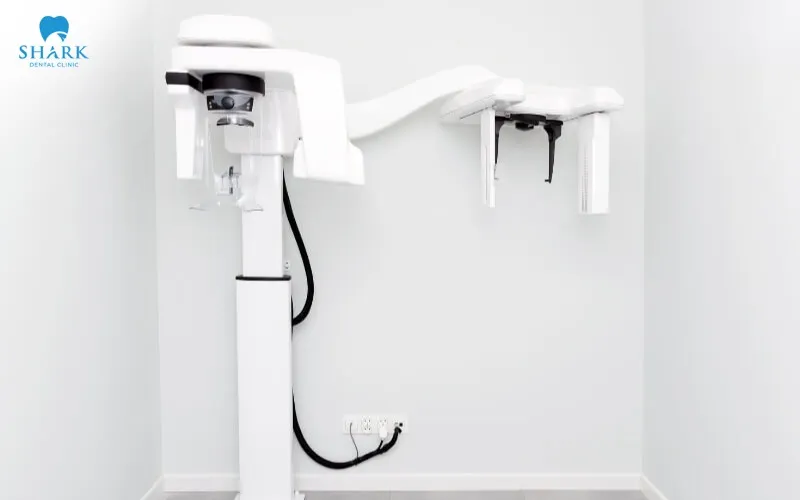
certain abnormalities that fall within the area being captured.
That said, since a panoramic film is a two-dimensional image, it can be subject to a certain degree of magnification or overlapping of structures. For this reason, a panoramic X-ray is not the only imaging tool available for every diagnostic situation; rather, it needs to be indicated by the dentist and combined with other suitable methods whenever this is necessary.
How does the panoramic X-ray machine work?
During the imaging process, the panoramic X-ray machine operates by having the X-ray-emitting head and the image-receiving component move together around the patient’s head. The resulting data is then processed by the system in order to create a panoramic image of the teeth, jaws, and surrounding structures in a single panoramic image.
In order for the resulting image to be clear and to help limit any inaccuracies, the patient needs to stand or sit in the correct position and to place their chin and bite onto the positioning device according to the instructions given. During the imaging process itself, the patient needs to keep their head steady, position their tongue correctly whenever instructed to do so, and remain still until the entire process of capturing the image has been completed.
What does a Panoramic X-ray machine film help to assess?
Thanks to its ability to capture an overview image of both dental arches in a single panoramic image, a Panoramic X-ray machine can help the dentist to observe many different areas and conditions relating to the patient’s teeth and gums.
- Wisdom teeth and impacted teeth: Helps with observing the direction of eruption, the position, and the general relationship of these teeth to the surrounding structures.
- The overall condition of the teeth: Provides an overview of the number of teeth present, the tooth roots, and any areas that may require further examination.
- The jawbone: Helps with observing the overall structure of the jawbone and with detecting any areas that may require a more in-depth assessment.
- Orthodontics: Can be combined with a cephalometric film, other images, and dental models, in order to support the assessment process.
- Implant placement: Provides an overview image of the jaw area, and in many cases, the dentist may also indicate a Cone Beam CT scan in addition, in order to examine the structure concerned in three-dimensional space.
Applications of the Panorama panoramic X-ray within dentistry
Thanks to its ability to provide an overview image of both dental arches and the related structures, a Panoramic X-ray machine can be used in many different examination and treatment situations within dentistry. Some of its applications include the following:
- General dental examination whenever there is a clinical need for one.
- Helping to assess wisdom teeth and impacted teeth before extraction.
- Supporting the orthodontic treatment process.
- Supporting root canal treatment in situations that require an overview image.
- Supporting the assessment and treatment of periodontal disease.
- Supporting restoration involving multiple teeth.
- Initial examination before implant placement.
- Monitoring certain conditions or areas of damage within the jaw and facial region.
The decision to indicate a Panoramic X-ray machine needs to be based on the information that is actually necessary for diagnosis and treatment. It should not be repeated simply because a check-up is due, if there is no genuine clinical need to do so.
The process of taking a Panorama panoramic X-ray
In order to obtain a clear image that covers the correct area to be examined, the process of taking a Panoramic X-ray machine is carried out according to the following steps:
- Step 1 – Checking the indication and any existing films: The dentist determines the reason for the X-ray, and the technician checks the imaging request along with any previous X-ray films, if these are available, in order to avoid any unnecessary repeat imaging.
- Step 2 – Removing any objects that could affect the image: The patient needs to remove glasses, jewelry, metal objects, or any removable dental appliances that could potentially affect the quality of the resulting film.
- Step 3 – Positioning: The patient’s head, chin, and bite are placed in the correct position, in accordance with the instructions given.
- Step 4 – Capturing the image: The patient remains still while the machine carries out the process of capturing the image.
- Step 5 – Checking the film: The patient’s position, the clarity of the image, and the coverage of the film are all checked after the imaging process has been completed.
- Step 6 – Reading and assessment: The dentist reads the film in combination with the results of the clinical examination and any other relevant data available.
Notable features of the panoramic X-ray machine
Thanks to its ability to capture an overview image of the jaw and facial area in a single panoramic image, a Panoramic X-ray machine offers a great deal of convenience throughout the process of examination and assessment. Some of the notable features of this particular technique include the following:
- It captures an image of both dental arches in a single panoramic image.
- The imaging time involved is relatively short.
- It helps with observing an overview of many different areas within the jaw and facial region.
- It has value for screening purposes and for initial treatment planning.
- The digital data produced is convenient for storage and for comparison purposes.
- It helps the dentist to identify areas that may require more detailed imaging and assessment.
How is a Panoramic X-ray machine different from a Cone Beam CT scan?
Both a Panoramic X-ray machine and a Cone Beam CT scan are used to help support diagnosis within dentistry, but they provide different types of image data. A Panoramic X-ray machine produces a two-dimensional image, which helps with observing an overview of the jaw and facial region, and it can be subject to the overlapping or magnification of structures. A Cone Beam CT scan, on the other hand, provides three-dimensional image data, which allows the structure concerned to be examined across a number of different planes, and it is generally indicated whenever a more detailed assessment is required.
The choice between a Panoramic X-ray machine and a Cone Beam CT scan needs to be based on the diagnostic question involved and on the information that is actually necessary for the treatment process. Not every single case requires the use of an imaging tool that is capable of capturing a three-dimensional image.
Is a Panorama panoramic X-ray safe?
Since a Panoramic X-ray machine makes use of X-rays, there is still a certain level of radiation exposure involved during the imaging process. For this reason, the equipment needs to be operated according to the correct procedure, and it should only be used whenever there is a genuine, appropriate professional need to do so.
Before the X-ray is taken, the patient needs to inform the dentist if they are currently pregnant or suspect that they may be pregnant, if they have recently had a dental X-ray taken, if they have any relevant older films available, or if they have any difficulty maintaining the required position. Based on this information, the dentist will weigh up the diagnostic benefit involved and choose whichever option is most suitable.
Limitations of the panoramic X-ray machine
Alongside its ability to provide an overview image, a Panoramic X-ray machine still has certain limitations when it comes to assessing the detailed structure of the teeth and the jawbone.
- It does not fully represent the structure concerned in three-dimensional space.
- It can be subject to the overlapping of structures within the image.
- It is not optimal for detecting every small area of damage.
- It does not replace a periapical film, a bitewing film, or a Cone Beam CT scan whenever a more detailed image is required.
- The quality of the resulting image can be affected by the patient’s position and by any movement that occurs during the imaging process.
Frequently asked questions
Each individual case involving a Panoramic X-ray machine may have a different purpose and different requirements. For this reason, when learning about this device, many patients still have questions about the process and the indications involved. Set out below are some of the most frequently asked questions.
Is taking a Panoramic X-ray machine painful?
The answer is no. The process of taking the X-ray does not cause any pain; the patient simply needs to stand or sit in the correct position as instructed, and remain still while the image is being captured.
Is a Panorama film enough on its own for implant placement?
Not every single case requires only a Panorama film to be taken. Depending on the specific condition involved, the dentist may also indicate a Cone Beam CT scan in addition, in order to assess the size of the bone and the anatomical relationships involved in three-dimensional space.
Can children have this type of X-ray taken?
Yes, it can be taken whenever there is a suitable indication to do so. The parameters and the coverage of the image need to be selected based on the diagnostic needs of each individual case.
The panoramic X-ray machine helps to capture an overview image of both dental arches and the related structures, and in doing so, it helps the dentist to identify any areas that may require further assessment and to draw up a suitable treatment plan. However, a Panorama film does not replace the need for a clinical examination or for specialized imaging methods whenever a more detailed examination is required. Shark Dental Clinic always places patient safety first, and it consistently invests in its systems and equipment in order to help deliver the results that patients expect. Let Shark Dental Clinic accompany you on your journey toward a new, more even, and more beautiful smile, every single day.