

The digital dental scanner is a piece of equipment that is widely applied within modern dentistry, and it is used to help record the structure of the teeth and gums in the form of 3D data. Thanks to its ability to reconstruct a visual model, this device helps to make things more convenient for the dentist throughout the course of examination, treatment planning, and restoration design. In order to gain a clearer understanding of this device, let us explore the details together with Shark Dental Clinic right away below.
What is a digital dental scanner?
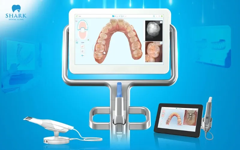
A digital dental scanner, which is also referred to as an intraoral scanner, is a piece of equipment that uses a small scanning tip in order to continuously record the surface of the teeth and the surface of the soft tissue within the area being observed. The resulting image data is then processed by software and reconstructed into a three-dimensional model, which is displayed directly on a screen.
This data can be used in place of a traditional dental impression in cases where doing so is considered suitable and appropriate. The dentist is fully able to enlarge the model in order to observe each individual area in much greater detail, and at the same time, the data can be transferred to the design department or to the laboratory without the need to physically transport a plaster model.
How does a digital dental scanner work?
The dentist moves the scanning tip in turn across the various surfaces of the teeth, so that the device is able to record image data and reconstruct it into a 3D digital model of each individual dental arch. The process of digital dental scanning typically follows the steps set out below:
- Step 1: The dentist begins by scanning the upper dental arch.
- Step 2: After this, the dentist continues on to scan the lower dental arch.
- Step 3: The patient bites the two arches together, following the dentist’s instructions, so that the dentist is able to record the relationship of the bite.
- Step 4: The dentist checks carefully for any areas of data that may still be missing, and carries out additional scanning of those areas if this is needed.
- Step 5: Once this whole process has been completed, the resulting data is stored in the form of a digital model.
The resulting model then serves as a valuable source of data that helps both the dentist and the dental technician throughout the entire process of designing restorations or other dental appliances. The device does not provide a diagnosis all on its own, and it does not replace the need for a full clinical examination.
What does the dental scanner help to support?
The digital dental scanner provides a source of 3D data that helps to support the process of examination, treatment planning, and restoration design. Specifically, this device fulfills the following roles:
- Recording the shape of the teeth: The 3D data shows the shape, the size, and the relative position of each tooth within the scanned area, which helps to support the process of assessing the overall structure of the teeth and gums.
- Providing a visual view: The image can be enlarged, rotated, and viewed from a number of different angles, which helps the dentist to explain the condition of the patient’s teeth and gums, as well as the treatment plan itself, in a way that is more visual and easier to understand.
- Supporting restoration design: The data that has been collected can be transferred into CAD/CAM software in order to design crowns, veneers, inlays, onlays, bridges, or implant-supported restorations.
- Storage and comparison: The digital model is convenient to store, and it can also be used for comparison purposes whenever it becomes necessary to track how the patient’s teeth change over the course of time.
Which services make use of the digital dental scanner?
The digital dental scanner is applied across many different areas of dentistry, and it helps to record the data that is needed in order to support the treatment process and to design restorations effectively. Some of the services that commonly make use of this technology include:
- Porcelain crowns and porcelain veneers.
- Inlays, onlays, and bridges.
- implant-supported restorations.
- Orthodontic treatment using braces or clear aligners.
- The design of retainers or night guards.
- Removable restorations in certain cases.
- Monitoring the shape of the teeth and the dental arch over time.
The extent to which the device can actually be applied will depend on the overall condition of the patient’s teeth and gums, on the particular area that needs to be recorded, and on the specific requirements of each individual type of restoration.
The digital dental scanning process at Shark Dental Clinic
The dental scanning process is carried out step by step, in order to fully record the data relating to the teeth, and this helps the process of treatment planning and restoration design to proceed more quickly. Specifically, the process typically includes the following steps:
- Step 1 – Examination: Before scanning takes place, the patient’s teeth, gums, and bite are checked by the dentist, and the specific purpose of taking the impression is determined.
- Step 2 – Preparation of the oral cavity: The surface of the teeth is cleaned, and saliva is controlled, before the scanning process begins.
- Step 3 – Scanning of the dental arch: The scanning tip is moved in turn around the surfaces of the teeth, following a sequence that is appropriate, in order to record the necessary data.
- Step 4 – Recording the bite: The patient bites the two arches together in the exact position that the dentist has instructed.
- Step 5 – Checking the data: The dentist checks the 3D model that has been produced and carries out additional scanning if this proves necessary.
- Step 6 – Saving and transferring the data: The data is either stored or transferred to design software and to the laboratory, so that it can be used for the following steps.
Notable features and limitations of the digital dental scanner
The digital dental scanner is a technology that is widely applied within dentistry. However, alongside its notable features, this device also has certain limitations that you should be aware of before deciding to go ahead with it.
Notable advantages
The digital dental scanner offers a number of notable advantages throughout the process of recording and processing data, including the following:
- Displaying a 3D model of the teeth directly on the screen: Once the scanning has taken place, the data is reconstructed into a 3D model, which is then displayed directly on the screen for the dentist to observe.
- Checking the data and carrying out additional scanning when needed: During the scanning process, the dentist is able to check the data that has already been recorded and carry out additional scanning of any areas that are still missing, whenever this is necessary.
- Convenience throughout the storage and transfer of data: The digital data can be stored and transferred to the design department or to the laboratory, without any need to physically transport an impression or a plaster model.
- Supporting a connection with the digital restoration design process: The scanned data can be used directly within the process of digital restoration design.
- Making it easy to visualize the condition of the teeth and the treatment plan: The visual 3D model makes it easy for the patient to see the condition of their own teeth, and this in turn allows the dentist to explain the situation and to draw up a suitable treatment plan.
Certain limitations
Alongside its notable advantages, the digital dental scanner is not always able to completely replace the traditional method of taking an impression. Recording data can prove difficult in certain situations, such as the following:
- The area to be scanned lies deep beneath the gum line: In this situation, the data may be more difficult to record than usual, since the area that needs to be scanned lies deep beneath the gum tissue.
- There is a large amount of saliva or bleeding present: The process of recording data can become more difficult when there is a large amount of saliva or bleeding present within the oral cavity.
- The patient has difficulty opening their mouth or staying still: The scanning process can be affected if the patient has difficulty opening their mouth wide enough, or has difficulty staying still in line with the dentist’s instructions.
- The area requiring restoration is particularly wide: In certain cases where the area requiring restoration is especially wide, recording the necessary data can become more difficult than usual.
- A long span of missing teeth with few anatomical landmarks: Recording data can be somewhat limited in cases where there is a long span of missing teeth and relatively few anatomical landmarks available for the software to use when comparing and stitching the data together.
Depending on each specific case that is involved, the dentist will carefully consider whether to use a digital impression, a traditional impression, or some combination of both methods, in order to best suit the treatment needs of that particular patient.
Technologies that are commonly combined with the digital dental scanner
In order to support the process of treatment planning and restoration design, the digital scanner is commonly combined with a number of the following technologies and systems:
- CAD/CAM systems and restoration design software.
- Cone Beam CT scanning and implant positioning systems.
- Orthodontic planning software.
- 3D printers or restoration fabrication machines.
Some frequently asked questions
While learning about this device, many patients tend to have a number of questions relating to how it is used, how it works, and the situations in which it should be carried out. Set out below are some of the most frequently asked questions.
Is dental scanning painful?
The scanning tip only moves across the surface of the teeth, and it does not emit any X-rays during the process of recording data. When scanning the teeth located further back in the mouth, the patient may feel a slight sensation of the tip being present, but in general, this method does not cause any pain.
Does dental scanning replace the need for an X-ray?
The answer is no. The scanner only records the surface of the teeth and the soft tissue within the area being observed, whereas an X-ray helps to assess internal structures such as the tooth roots and the jawbone.
Is it possible to carry out scanning in every single case?
It is not the case that every single situation is suitable for digital dental scanning. The dentist will carefully consider this, based on the actual condition involved and on the requirements of each particular type of restoration.
In summary, the digital dental scanner helps to record the surface of the teeth, the soft tissue within the area being observed, and the relationship of the bite, all in the form of 3D data. This data helps the dentist throughout the process of assessment, treatment planning, discussion with the patient, and the design of restorations or other dental appliances. However, the device does not replace the need for a clinical examination, an X-ray, or any other necessary diagnostic methods.
Shark Dental Clinic applies the digital dental scanner in cases where it is considered suitable, in order to help make the process of recording and processing data more convenient. Contact the hotline at 1800 2069 so that a dentist can examine you and advise on a suitable treatment option.