

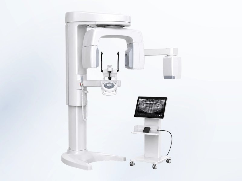
The 3D Cone Beam CT machine is a diagnostic imaging device that is applied within dentistry whenever the dentist needs to examine the structure of the teeth, jaws, and face in three-dimensional space. The resulting data helps the dentist with observing the teeth, the jawbone, and the relationships that exist between the various anatomical structures that fall within the particular area being scanned. A Cone Beam CT scan needs to be indicated by the dentist based on the diagnostic need involved and on the treatment plan that has been drawn up for each individual case. In the article set out below, Shark Dental Clinic will provide you with detailed information about this particular piece of equipment.
What is a 3D Cone Beam CT machine?
Cone Beam CT, which is also known as cone-beam computed tomography, or CBCT, is an imaging technique that uses X-rays in order to capture a number of projections around the head and facial region. This resulting data is then processed carefully by software and reconstructed into a full three-dimensional image.
The dentist is fully able to examine the data across a number of different planes, in order to carefully assess the structures that fall within the scanned area. When compared with a two-dimensional Panoramic X-ray film, CBCT provides additional information about the width, the height, and the depth of the area being examined, while also helping to limit the effect of any overlapping images.
That being said, it should be noted that a Cone Beam CT scan is not a necessary imaging tool for every single case that comes through the clinic. Because the device makes use of ionizing radiation, the scan is only ever indicated whenever the resulting information carries genuine, meaningful value for the diagnostic or treatment process in question.
How does a Cone Beam CT machine work?
During the course of the scanning process, the X-ray-emitting component and the image detector move together around the patient’s head, in order to capture a number of projections from a range of different angles. This data is then processed by software and reconstructed into a three-dimensional image. The scanning process typically consists of the following steps:
- Step 1 – Removing any metal objects: Any metal objects that could potentially affect the quality of the resulting image are removed whenever this is requested by the technician.
- Step 2 – Standing or sitting in the correct position: The patient follows the instructions given by the dentist, so that the device is able to properly record data within the area that needs to be examined.
- Step 3 – Keeping the head steady: The patient maintains their position carefully and limits any unnecessary movement throughout the entire scanning process.
- Step 4 – Not moving or swallowing: The patient remains completely still while the device is in the process of recording data.
The quality of the resulting data can be affected by a number of factors, including movement, metal materials present in the mouth, the particular area being scanned, and the technical parameters that have been selected for the scan.
What does a Cone Beam CT machine help to assess?
A Cone Beam CT machine provides three-dimensional image data, which helps the dentist to examine the teeth, the jawbone, and the various anatomical structures that fall within the area being scanned. Some of the situations in which this scan may be indicated include the following:
- Assessing the jawbone: Examining the size, the shape, and the structure of the bone within the scanned area.
- Implant placement: Helping to assess the area where an implant is expected to be placed, along with its relationship to any anatomical structures that need to be taken into account.
- Wisdom teeth and impacted teeth: Observing the position, the direction of eruption, and the three-dimensional relationship of the tooth to the surrounding structures.
- Endodontic treatment: Helping to examine the root canal system, any periapical lesions, or any complex situations in which two-dimensional imaging has not provided sufficient information.
- Oral and maxillofacial surgery: Helping to assess the area requiring intervention and to plan the surgical procedure.
- Orthodontics: Providing three-dimensional data in cases where an assessment of the craniofacial structure, impacted teeth, or the airway is indicated.
- Conditions affecting the jaw and facial region: Helping to examine certain bone lesions, cysts, or abnormalities that fall within the scanned area.
CBCT is a diagnostic support tool; it does not determine a diagnosis all on its own, and it does not replace the need for a proper clinical examination carried out by the dentist.
Which services make use of the Cone Beam CT machine?
The Cone Beam CT machine can be applied across many different dental services whenever the dentist needs additional three-dimensional image data in order to support diagnosis and treatment planning.
- Single-tooth or full-arch implant placement.
- Sinus lifting and bone grafting.
- Extraction of wisdom teeth or impacted teeth.
- Endodontic treatment in cases that require a three-dimensional examination.
- Orthodontic treatment for which a CBCT scan is indicated.
- Oral and maxillofacial surgery.
- Examination of certain lesions affecting the teeth and the jawbone.
- Treatment planning supported by implant positioning technology.
It is not the case that every single situation falling within the categories listed above necessarily requires a CBCT scan to be carried out. The dentist will make this particular decision based on the information that is actually needed for the diagnostic and treatment process involved in each individual case.
The process of taking a Cone Beam CT scan at Shark Dental Clinic
The process of taking a Cone Beam CT scan at Shark Dental Clinic is carried out step by step, in order to record image data that is appropriate to the diagnostic goal involved:
- Step 1 – Examination and determining the indication: The dentist assesses the clinical condition, any existing films, and the information that needs to be examined, in order to arrive at an appropriate indication for the scan.
- Step 2 – Preparation before the scan: The patient removes their glasses, jewelry, removable dental appliances, or any metal objects that could potentially affect the image data.
- Step 3 – Selecting the scanning area: The area to be scanned and the parameters of the scan are both selected in a way that is appropriate to the diagnostic goal of each individual case.
- Step 4 – Positioning: The patient’s head and maxillofacial region are placed in the correct position, in accordance with the instructions given, so that the device is able to record the data.
- Step 5 – Capturing the image: The patient remains still while the device rotates around the head area and captures the image.
- Step 6 – Checking the data: The technician checks the quality and the coverage of the image to make sure that the data meets the necessary requirements.
- Step 7 – Reading and assessment: The dentist analyzes the CBCT data in combination with the clinical examination and any other relevant information, in order to support the diagnostic and treatment process.
Notable features of the 3D Cone Beam CT machine
The 3D Cone Beam CT machine provides image data in three-dimensional space, helping the dentist to examine and analyze the structures that fall within the scanned area. Some of its notable features include the following:
- Providing three-dimensional image data: Allows the structures within the scanned area to be examined in 3D space.
- Examination across multiple planes and viewing angles: Helps with observing structures from a number of different directions.
- Supporting measurement and treatment planning: The data can be used within software in order to carry out measurements and to support treatment planning.
- Limiting the effect of overlapping images: Helps to reduce the effect of the overlapping that is commonly seen on two-dimensional films.
- Convenient for storage and comparison: The digital data can be stored and used for comparison purposes whenever this is needed.
- Can be combined with treatment software and systems: The data can be combined with implant planning software and with surgical positioning systems.
The accuracy of any measurements that are taken, and of the way in which the data is ultimately applied, still depends on a range of factors, including the equipment used, the scanning parameters selected, the patient’s own movement, any image artifacts that may occur, and the analysis carried out by an appropriately qualified professional.
How is Cone Beam CT different from a Panoramic X-ray?
| Criteria | Panoramic X-ray | Cone Beam CT |
| Data | Two-dimensional image | Three-dimensional data |
| Purpose | Overview of both dental arches and related structures | Detailed examination across multiple planes |
| Overlapping | Can occur | More limited, thanks to three-dimensional data |
| Measurement | Can be affected by magnification | Supports three-dimensional measurement within software |
| Radiation | Generally lower than CBCT | Varies according to the equipment, the area, and the parameters used |
| Indication | Whenever an overview image is needed | Whenever three-dimensional data adds diagnostic value |
Cone Beam CT should never simply be assumed to be better than a Panoramic X-ray in every single situation that might arise. The most suitable imaging tool is the one that provides sufficient information for the diagnostic and treatment goal involved, while also ensuring a reasonable level of exposure.
Is Cone Beam CT safe?
Since Cone Beam CT makes use of X-rays, there is still a certain level of radiation exposure involved. When there is an appropriate professional indication, the diagnostic benefit can outweigh the relatively low level of radiation risk involved. That said, the scan needs to follow the principle of selecting the correct indication and of using a scanning area and parameters that are appropriate to the information being examined. Before the scan is taken, the patient should inform the dentist if they:
- Are currently pregnant or suspect that they may be pregnant.
- Have recently had a dental X-ray or CT scan taken.
- Have any related films or diagnostic imaging data available.
- Have difficulty standing, sitting, or remaining still.
- Have metal materials or removable devices within the head and facial region.
For children specifically, the decision to carry out a CBCT scan needs to be very carefully considered, and the parameters that are selected need to be appropriate to the child’s particular age, their body size, and the diagnostic goal that is involved in each case.
Limitations of the Cone Beam CT machine
Alongside its ability to provide three-dimensional imaging, the Cone Beam CT machine still has certain limitations that should be kept in mind:
- It makes use of ionizing radiation.
- It should not be used as a routine screening tool in the absence of an appropriate professional indication.
- The resulting image can be affected by metal objects, movement, or incorrect positioning during the scanning process.
- Its ability to distinguish between certain types of soft tissue remains more limited than that of specialized soft-tissue imaging tools that are designed specifically for that purpose.
- It does not replace the need for a clinical examination and assessment.
- Reading and assessing the entire set of data that is produced needs to be carried out by an appropriately qualified professional with the right expertise.
- It may reveal findings that fall outside the original purpose of the scan, and these particular findings may need further assessment whenever this proves to be necessary.
Which technologies is Cone Beam CT commonly combined with?
Cone Beam CT can be combined with a number of different digital technologies and systems within dentistry, helping to support the overall process of diagnosis, planning, and treatment.
- Digital dental scanner: Combines 3D image data with tooth surface data in cases where this is appropriate.
- Implant planning software: Helps with planning based on the Cone Beam CT data.
- Implant positioning systems: Combines image data in order to support the positioning process.
- CAD/CAM systems: Helps with processing digital data throughout the design and fabrication process.
- Orthodontic and surgical planning software: Provides data to support appropriate planning processes.
- 3D printers or surgical guide fabrication systems: The data can be used within the process of designing and fabricating a surgical guide.
Frequently asked questions
Cone Beam CT provides three-dimensional imaging and is applied across a wide range of different dental situations. That said, patients may still have a number of questions about this device; set out below are some of the most frequently asked questions.
Is a Cone Beam CT scan painful?
Not at all. The scanning process does not cause any pain. The patient simply needs to stand or sit in the correct position and remain still while the device records the data.
Does a Cone Beam CT scan require a contrast agent to be injected?
In typical dental applications, CBCT generally does not make use of a contrast agent. If a specialized assessment of soft tissue or blood vessels is required, the dentist will select an appropriate imaging tool for that purpose.
Is a Cone Beam CT scan mandatory for implant placement?
The indication for a CBCT scan depends on the patient’s condition and on the information that is needed for the treatment plan. In many cases involving implant placement, CBCT provides valuable three-dimensional data for assessing the bone and the anatomical structures involved. That said, the dentist still needs to make this decision based on each individual case.
Does Cone Beam CT replace all other types of dental X-rays?
The answer is no. Periapical films, bitewing films, panoramic radiographs, and CBCT each have a different scope of application. The dentist will select whichever technique is capable of providing sufficient information for the diagnostic question involved, while also avoiding any unnecessary imaging tests.
The 3D Cone Beam CT machine provides three-dimensional image data of the dental and maxillofacial region, helping the dentist to assess anatomical structures and to draw up a treatment plan in cases where this is appropriate. The device does not determine a diagnosis on its own, and it is not a method that is necessary for every single patient. At Shark Dental Clinic, the decision to take a Cone Beam CT scan is carefully considered based on the diagnostic need and the treatment plan of each individual case. Contact the hotline at (+84) 941 623 322 to arrange an examination and receive suitable advice from a dentist.